
Dr Olivier Peltre (Post-Doctoral Researcher) and Dr Yaël Frégier (Senior Lecturer)
“The team here at the University of Artois are using neural networks to identify relevant features in intracranial pressure (ICP) signals and flow signals (arterial and venous blood flow, and cerebrospinal fluid (CSF) flow).
The first step is to collect, anonymise and prepare the data captured over the past 20 years by our partners in Cambridge and Amiens: several thousands of datasets (infusion test and phase contrast MRI) have been collected in this way. Moving forward, REVERT will begin collecting data combining these two examinations for the first time.
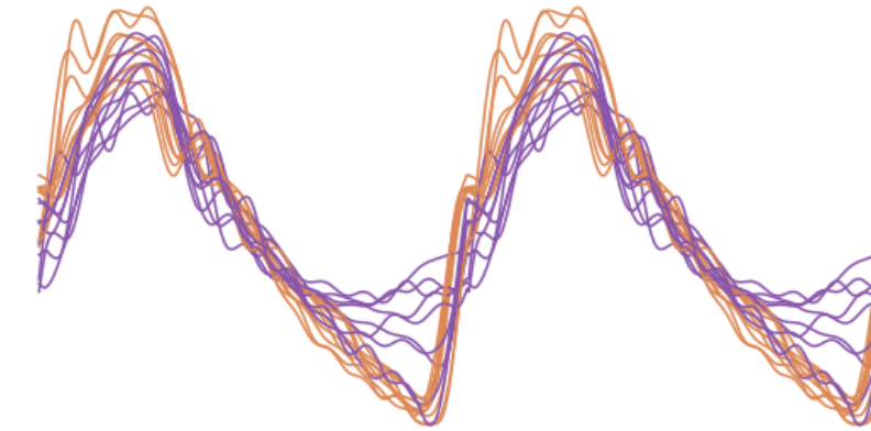
The PC-MRI sequence developed in Amiens records a dozen blood and CSF pulses in parallel over the duration of a typical cardiac cycle, corresponding to the regions of interest selected by the radiologist (carotid artery, jugular vein, CSF moving through the cervical vertebrae, etc.). An infusion test enables high-resolution monitoring of intracranial pressure over a period of 30–60 minutes, amounting to several thousand pulses per patient. In order to compare these two sets of data, we have started segmenting the ICP signal to extract a set of unique, representative pulses linked to each patient.
One benefit of having several thousand pulses per patient is that we will be able to train a neural network to identify whether two given pulses come from the same patient. This example of a ‘pretext task’ is at the heart of our self-supervised learning strategy, which will enable us to get the most out of the data.

What is unique and new about the REVERT project is that it brings together these two examinations (infusion test and PC-MRI) within the patient’s clinical pathway, and will provide a comprehensive insight into the cerebrospinal system (showing both pressure and volumetric flows), thus enabling a more accurate diagnosis of normal pressure hydrocephalus.”